What is Q fever?
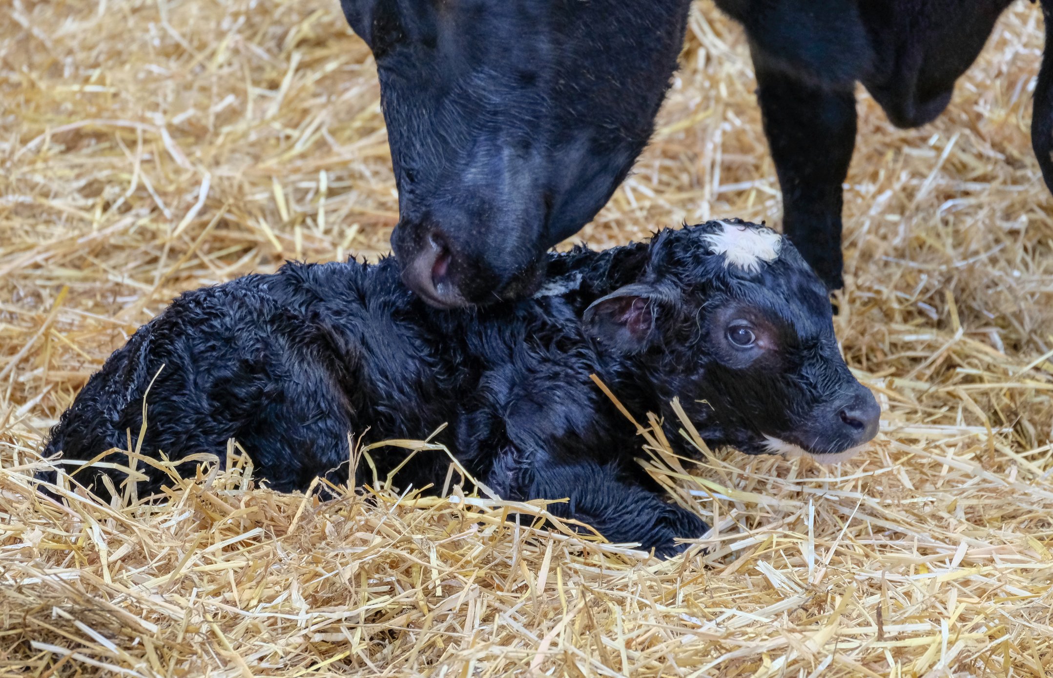
Q fever is a highly contagious zoonosis, that also has a negative impact on the health and reproductive performance of ruminants. Signs in cattle may include abortion, premature, still born or weak calves and infertility.
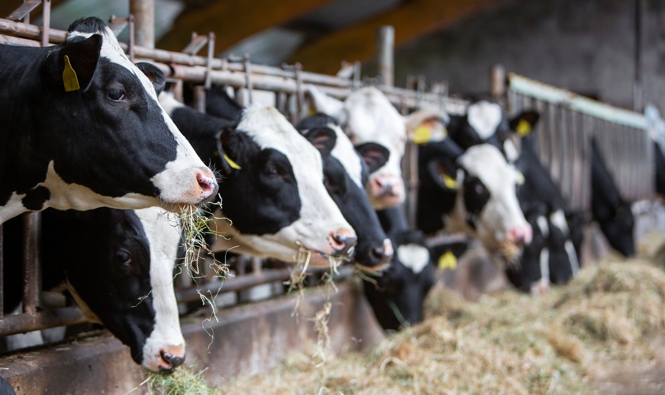
There is a varying prevalence reported in the UK and Ireland1,2,3,4 which includes 80% prevalence of positive Bulk Milk (BM) ELISA across 225 GB dairy herds and 70% BM PCR positive in South West England.
However, as the disease is mostly insidious and the diagnosis can be quite challenging, only a small percentage of farmers know that Q fever is present on their farms.
Learn more about this disease on our Q fever website.

How a farm becomes infected
Q fever is caused by the bacterium Coxiella burnetti, which is highly infectious and highly contagious. The bacteria, due to its small size, can travel up to 11 miles with the wind, transmitting the disease from an infected farm to a healthy one.
Additionally, a farm can become infected through the introduction of an animal already infected with C. burnetii. This infected animal, through shedding of the bacteria in vaginal discharge, parturition fluids and faeces, will contaminate the environment and then, via airborne spread, transmit the disease to healthy animals.
When to suspect Q Fever?
The main Q Fever symptoms in ruminants are reproductive problems, including abortions (including early embryo loss or reabsorptions), stillbirths, and infertility. A recent and unexplained degradation of the reproductive performances of the herd that manifest with an elevated number of retained placenta, metritis difficult to treat and poor fertility certainly warrants a Q fever diagnostic investigation. In goats and sheep, acute Q fever will often manifest as the main causes of abortion storms.
How can I confirm that a herd is infected?
A diagnosis of Q Fever can be a challenge for several reasons including:
- Infected animals can be asymptomatic but still be shedding the bacteria
- Animals can shed from differing routes and the amount of shedding can vary significantly over time
- In cattle the presentation is often subclinical and can be confused/diluted by other factors
In an effort to simplify the diagnosis of this disease, Ceva’s ruminant team has developed a new PCR diagnostic tool for Q fever, the Q Test. This is validated for use on bulk tank milk and is available through vets from Ceva.
Serology ELISAs are available to measure antibody levels, which can be useful as an indicator of exposure to the bacteria in unvaccinated animals.
To find out about Ceva supported Q fever diagnostics please contact us.

Prevention is key
Q Fever vaccination is important for protecting herds, by decreasing shedding in infected herds to reduce the clinical impact of the disease, reduce zoonotic risk and break the cycle of the bacteria.
Coxevac has proven efficacy in reducing shedding of the bacteria by ruminants. By controlling the disease in animals, the vaccine also reduces the risk of transmission to humans.

Q fever heat map
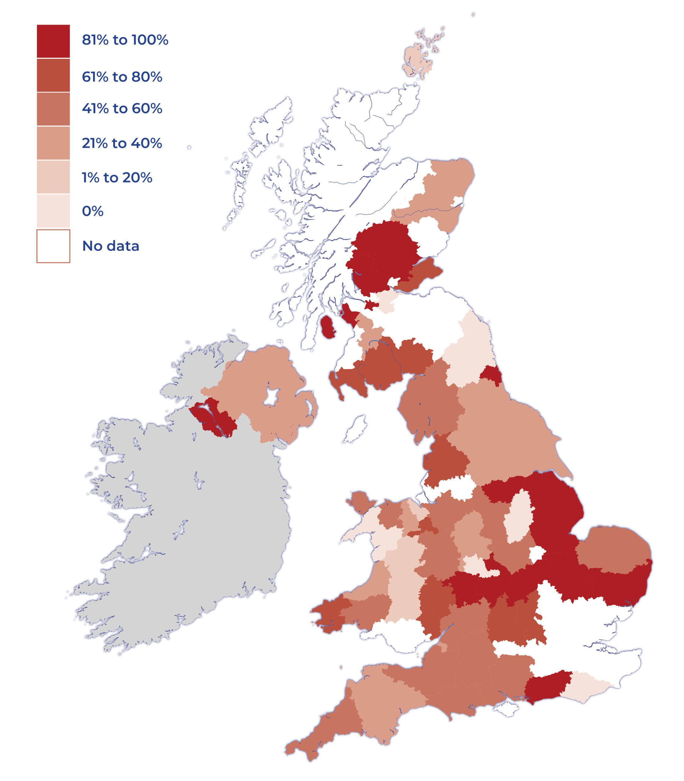
Incidence of Q fever in UK dairy herd as diagnosed by Q Test (PCR)
- Total number of Q tests reported to March 2026: 1000
- National average 45% positive
- Recording of data commenced 2020